
Michigan State University · Department of Radiology
Seeing the human brain in unprecedented detail
CIRC is MSU's hub for human neuroimaging — integrating multi-modal imaging with next-generation analysis to decode the brain in health and disease.
Our mission
Bridging image acquisition and analysis
CIRC brings together investigators from across MSU to turn state-of-the-art imaging into insight — improving lives today and tomorrow.
Research in action
From raw signal to brain biomarker
CIRC investigators map the brain's structure, function, and metabolism — resting-state networks, BOLD activation, diffusion tractography, and simultaneous PET/MR — and translate those signals into biomarkers of cognition, aging, and disease.
Imaging modalities
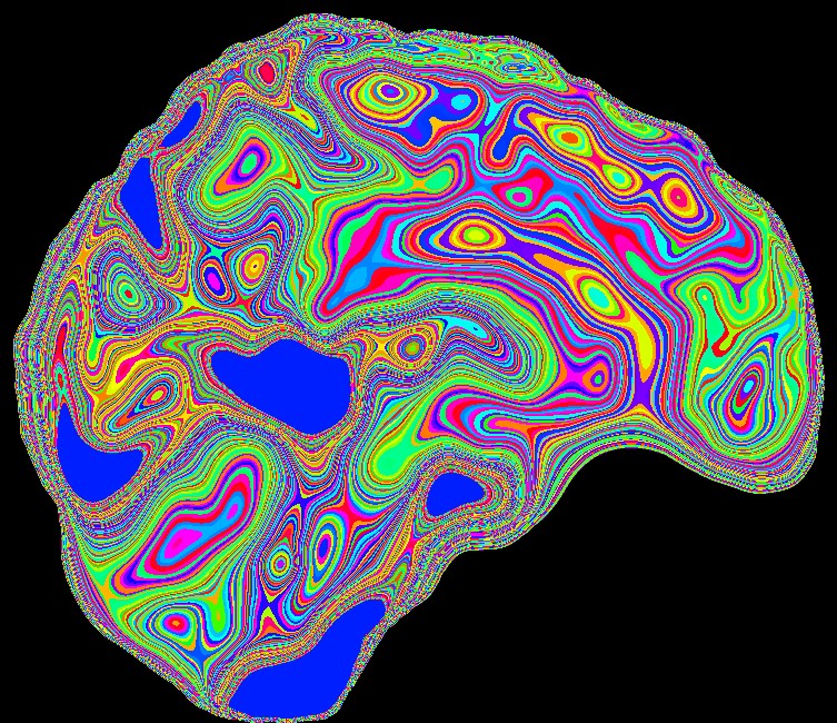
Many ways to see the brain
Structure, function, connectivity, and metabolism — CIRC investigators image the brain across modalities and fuse them into a single picture of brain health.
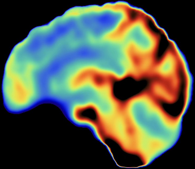
Resting-state function
Spontaneous BOLD fluctuations (ALFF) mapping the brain's intrinsic activity.
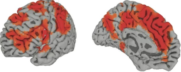
BOLD activation
Task-evoked responses rendered on inflated cortical surfaces.
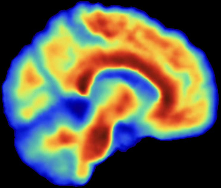
Positron Emission Tomography
Molecular imaging with amyloid, tau, and FDG PET on the Biograph One PET/MR.
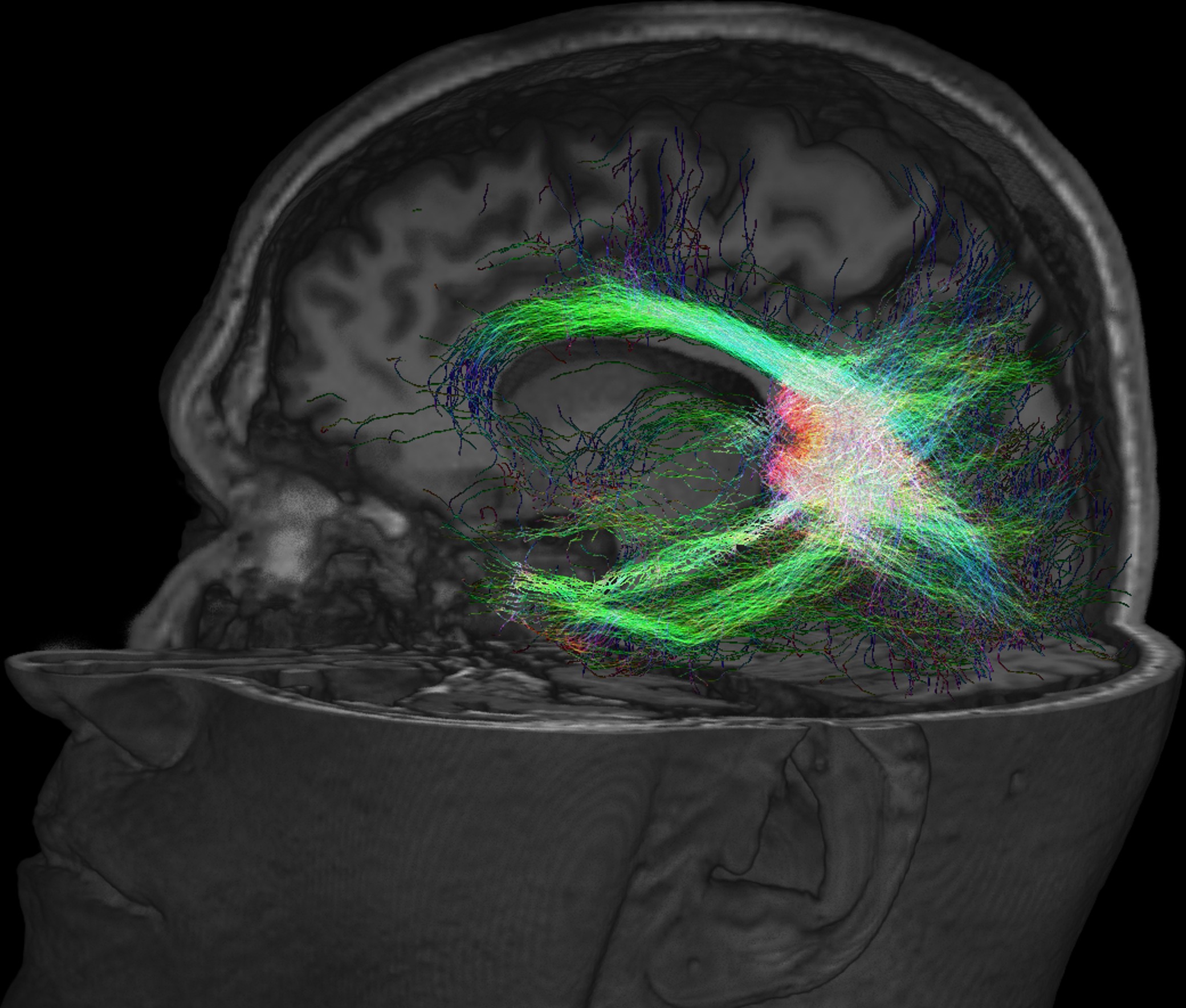
Diffusion tractography
White-matter pathways reconstructed from diffusion MRI.
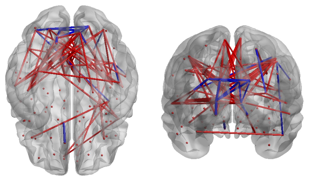
Functional connectivity
Networks that reveal how distant brain regions communicate.
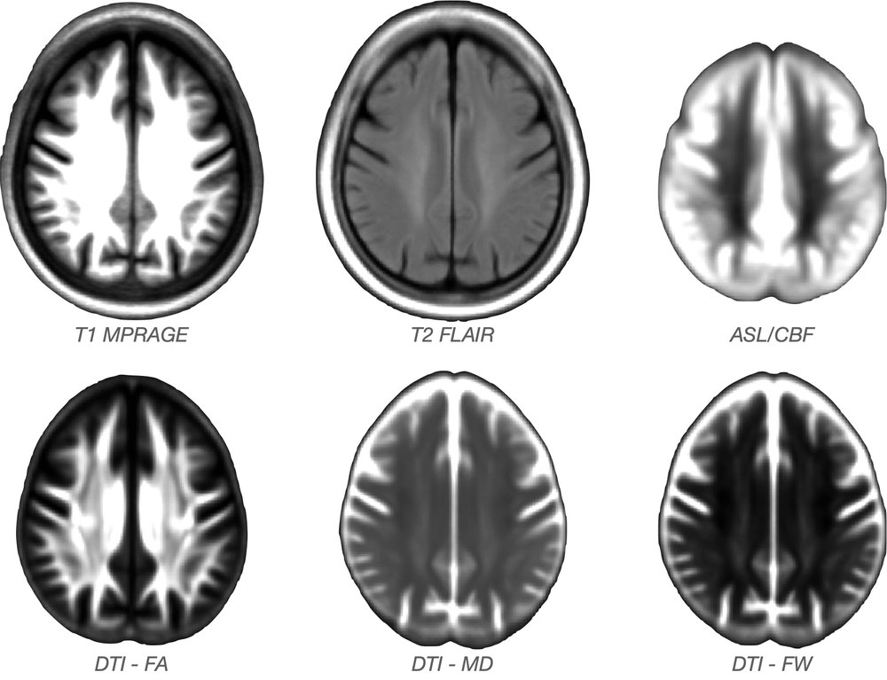
Multi-modal templates
T1, FLAIR, ASL perfusion and diffusion — harmonized in one space.
The brain in motion
Function you can watch unfold
Beyond static maps, our analyses capture the brain's dynamics — the moment-to-moment ebb and flow of activity across the cortex. These signals help us turn raw fluctuations into biomarkers of cognition, aging, and recovery.
Now operational
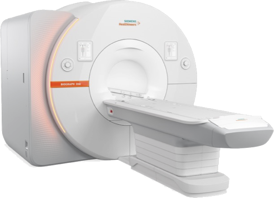
Siemens Biograph One PET/MR
CIRC is a proud partner of the Center for Imaging and Image-Guided Therapies (CIIGT) — pronounced "see-get" (See the image, Get the result). Housed within this facility, our Biograph One is one of the first systems of its kind installed in the United States.
Simultaneous capture of metabolic and functional processes opens transformative avenues for research into cognition and neurodegeneration.
Partner with CIRC
Interested in scan time, collaboration, or facility access? We welcome investigators from across MSU and partner institutions.