
Facilities
World-class imaging & computing
CIRC investigators access a comprehensive suite of state-of-the-art imaging systems across MSU's East Lansing and Grand Rapids campuses and partner institutions — backed by high-performance computing and dedicated imaging scientists.
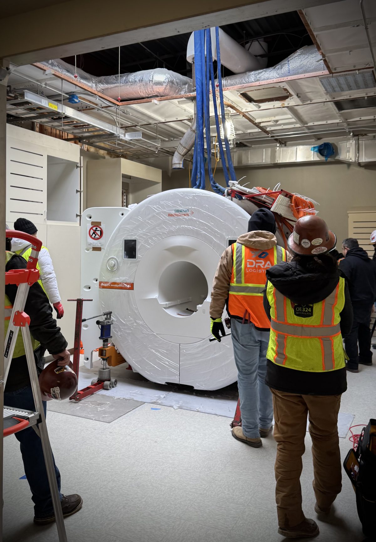
Siemens Biograph ONE 3T PET/MR
Located within the Center for Imaging and Image-Guided Therapies (CIIGT) at MSU Radiology, the Biograph One is a next-generation system for simultaneous PET and 3T MR acquisition — one of the first systems of its kind installed in the United States.
East Lansing campus
Additional MRI on the main campus
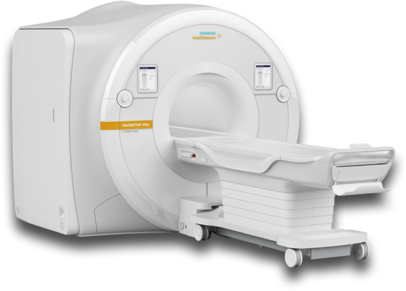
Siemens MAGNETOM Vida 3T
Accessed via MSU Health Care at the McLaren Greater Lansing Outpatient Imaging Center (Izzo Building). XT gradients (60 mT/m, 200 T/m/s), BioMatrix technology, and a comprehensive coil suite including 20- and 64-channel head coils.
SMS-accelerated fMRI and dMRI (DTI to NODDI), SWI, TWIST MRA, and MRS — with Deep Resolve Discover AI acceleration.

MSU Department of Radiology
Beyond the research systems, the Department operates a full range of clinical imaging — 4 clinical MRI scanners (2× 3T Vida, 2× 1.5T Sola), PET-CT, a radiopharmacy with two cyclotrons, CT, mammography, ultrasound, fluoroscopy, DEXA, and interventional services.
Grand Rapids campus · via BAMF Health & Corewell
Molecular imaging & total-body PET
On the Grand Rapids Medical Mile, CIRC investigators reach some of the most advanced molecular-imaging instrumentation in the country.
 BAMF Health
United Imaging uPMR 790 HD TOF PET/MR
Simultaneous 3T PET/MR with digital SiPM + LYSO detectors, excellent Time-of-Flight performance, 2.8 mm NEMA PET resolution across a 32 cm axial FOV, and the uCS accelerated-imaging platform.
 BAMF Health
United Imaging uEXPLORER Total-Body PET/CT
A 194 cm axial field-of-view images the entire body in a single acquisition — ~40× effective sensitivity, ultra-low-dose and sub-minute protocols, and total-body dynamic pharmacokinetics, with a 160-slice CT.
 Corewell Health
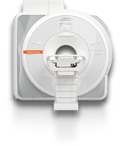
Siemens MAGNETOM Prisma 3T
Research-optimized 3T with ultra-high-performance XR gradients (80 mT/m, 200 T/m/s) for HARDI/DSI diffusion, high-resolution fMRI, advanced and multinuclear (³¹P) MRS, and Human Connectome Project protocols.
External collaborative facilities · via MADRC
Statewide imaging network

2× GE MR750 3T — University of Michigan fMRI Laboratory
High-performance gradients (50 mT/m, 200 T/m/s), 32-channel arrays, an extensive library of rapid pulse sequences (spiral, ASL, multi-band), sequence-compiler access, and MRS including GABA detection.
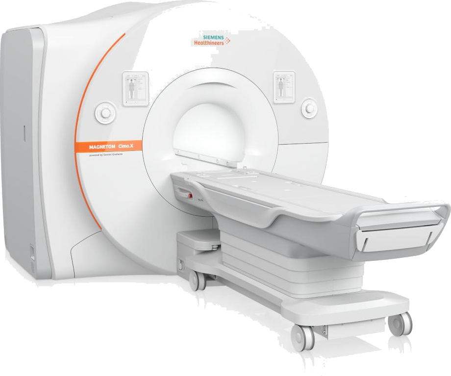
Siemens MAGNETOM Cima.X 3T — Wayne State MR Core
Newly installed, with revolutionary Gemini Gradients (200 mT/m at 200 T/m/s), next-generation Deep Resolve AI reconstruction, Open Recon, and 64-channel head/neck coils for advanced diffusion, fMRI, ASL, and multinuclear spectroscopy.
Stimulus, response & data
Stimulus presentation
MR-compatible projection with a high-resolution in-bore display. Paradigms are built and run predominantly in PsychoPy and the Psychtoolbox for MATLAB, synchronized to scan timing.
Response & monitoring
Fiber-optic button response systems and high-resolution eye tracking (EyeLink 1000 Plus) for accurate behavioral measurement during imaging.
Data & export
CIRC runs its own XNAT server for direct export from the scanner and self-service data download by investigators. Reach out to get set up.
Computation
From workstation to supercomputer
Analysis moves seamlessly from investigator workstations to MSU's high-performance computing center, so studies scale from a single subject to large, multi-site cohorts without changing tools.
For large-scale jobs, investigators use MSU's Institute for Cyber-Enabled Research (ICER) HPCC — with containerized pipelines, secure storage, and expert consulting. Neuroimaging runs on FSL, SPM, AFNI, MRtrix, FreeSurfer, and ANTs, plus in-house GPU-accelerated Python/MATLAB pipelines and Singularity containers.
MSU Department of Radiology — home of CIRC and the CIIGT imaging core, in the Biomedical Research Complex, East Lansing.